Showing posts with label Long QT. Show all posts

Thursday, February 1, 2018

Normal Impulse Conduction

Sequence of Cardiac Electrical Activation

conduction velocities in the heart
The action potentials generated by the SA node spread throughout the atria primarily by cell-to-cell conduction at a velocity of about 0.5 m/sec. There is some functional evidence for the existence of specialized conducting pathways within the atria (termed internodal tracts), although this is controversial. As the wave of action potentials depolarizes the atrial muscle, the cardiomyocytes contract by a process termed excitation-contraction coupling.
Normally, the only pathway available for action potentials to enter the ventricles is through a specialized region of cells (atrioventricular node, or AV node) located in the inferior-posterior region of the interatrial septum. The AV node is a highly specialized conducting tissue (cardiac, not neural in origin) that slows the impulse conduction considerably (to about 0.05 m/sec) thereby allowing sufficient time for complete atrial depolarization and contraction (systole) prior to ventricular depolarization and contraction.

heart depolarization times
The impulses then enter the base of the ventricle at the Bundle of His and then follow the left and right bundle branches along the interventricular septum.  These specialized fibers conduct the impulses at a very rapid velocity (about 2 m/sec).  The bundle branches then divide into an extensive system of Purkinje fibers that conduct the impulses at high velocity (about 4 m/sec) throughout the ventricles. This results in rapid depolarization of ventricular myocytes throughout both ventricles.

The conduction system within the heart is very important because it permits a rapid and organized depolarization of ventricular myocytes that is necessary for the efficient generation of pressure during systole. The time (in seconds) to activate the different regions of the heart are shown in the figure to the right. Atrial activation is complete within about 0.09 sec (90 msec) following SA nodal firing. After a delay at the AV node, the septum becomes activated (0.16 sec). All the ventricular mass is activated by about 0.23 sec.

Regulation of Conduction


The conduction of electrical impulses throughout the heart, and particularly in the specialized conduction system, is influenced by autonomic nerve activity. This autonomic control is most apparent at the AV node. Sympathetic activation increases conduction velocity in the AV node by increasing the rate of depolarization (increasing slope of phase 0) of the action potentials. This leads to more rapid depolarization of adjacent cells, which leads to a more rapid conduction of action potentials (positive dromotropy). Sympathetic activation of the AV node reduces the normal delay of conduction through the AV node, thereby reducing the time between atrial and ventricular contraction. The increase in AV nodal conduction velocity can be seen as a decrease in the P-R interval of the electrocardiogram.


sympathetic and vagal effcts on atrioventricular node action potentials

Sympathetic nerves exert their actions on the AV node by releasing the neurotransmitter norepinephrine that binds to beta-adrenoceptors, leading to an increase in intracellular cAMP. Therefore, drugs that block beta-adrenoceptors (beta-blockers) decrease conduction velocity and can produce AV block.


Parasympathetic (vagal) activation decreases conduction velocity (negative dromotropy) at the AV node by decreasing the slope of phase 0 of the nodal action potentials. This leads to slower depolarization of adjacent cells, and reduced velocity of conduction. Acetylcholine, released by the vagus nerve, binds to cardiac muscarinic receptors, which decreases intracellular cAMP. Excessive vagal activation can produce AV block. Drugs such as digitalis, which increase vagal activity to the heart, are sometimes used to reduce AV nodal conduction in patients that have atrial flutter or fibrillation. These atrial arrhythmias lead to excessive ventricular rate (tachycardia) that can be suppressed by partially blocking impulses being conducted through the AV node.
Phase 0 of action potentials at the AV node is not dependent on fast sodium channels as in non-nodal tissue, but instead is generated by the entry of calcium into the cell through slow-inward, L-type calcium channels. Blocking these channels with a calcium-channel blocker such as verapamil or diltiazem reduces the conduction velocity of impulses through the AV node and can produce AV block.
Because conduction velocity depends on the rate of tissue depolarization, which is related to the slope of phase 0 of the action potential, conditions (or drugs) that alter phase 0 will affect conduction velocity.  For example, conduction can be altered by changes in membrane potential, which can occur during myocardial ischemia and hypoxia. In non-nodal cardiac tissue, cellular hypoxia leads to membrane depolarization, inhibition of fast Na+ channels, a decrease in the slope of phase 0, and a decrease in action potential amplitude. These membrane changes result in a decrease in speed by which action potentials are conducted within the heart. This can have a number of consequences. First, activation of the heart will be delayed, and in some cases, the sequence of activation will be altered. This can seriously impair ventricular pressure development. Second, damage to the conducting system can precipitate tachyarrhythmias by reentry mechanisms.

Tuesday, January 9, 2018

Long QT


Long QT Syndrome

What is LQTS?

  • Long Q-T syndrome is a disorder of the heart’s electrical system.
  • The electrical activity of the heart is produced by the flow of ions (electrically charged particles of sodium, calcium, potassium, and chloride) in and out of the cells of the heart. Tiny ion channels control this flow.
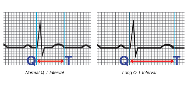
  • The Q-T interval is the section on the electrocardiogram (ECG) - that represents the time it takes for the electrical system to fire an impulse through the ventricles and then recharge. It is translated to the time it takes for the heart muscle to contract and then recover.
  • LQTS occurs as the result of a defect in the ion channels, causing a delay in the time it takes for the electrical system to recharge after each heartbeat. When the Q-T interval is longer than normal, it increases the risk for torsade de pointes, a life-threatening form of ventricular tachycardia.
  • LQTS is rare. The prevalence is about 1 in 5,000 persons in the United States.
Arrhythmia: Long Q-T Syndrome

What causes LQTS?

Long Q-T syndrome can be acquired or congenital:
Acquired LQTS is caused by many medications. Sensitivity to these medications may be related to genetic causes.
Congenital LQTS is usually inherited. It is caused by an abnormality in the gene code for the ion channels. The abnormality of the ion channels slows the recovery phase of the heartbeat. Forms of inherited LQTS include:
  • Recent Classifications – Multiple ion channel abnormalities have been discovered. The most common ones include LQT1, LQT2, LQT3, LQT4, LQT5; these are classified by the type of channel which causes the LQTS. The type of LQTS classification is related to the risk of future cardiac events, those with LQT3 having the highest risk of life-threatening arrhythmias.
  • Jervell, Lange-Nielsen Syndrome (autosomal recessive inheritance pattern) – Both parents are carriers of the abnormal gene, but they may not manifest LQTS. Each child has a 25-percent chance of inheriting LQTS. This syndrome is associated with deafness at birth and is extremely rare, as there is a small chance that both parents would carry the LQTS gene.
  • Romano-Ward Syndrome (autosomal dominant inheritance pattern) – One parent has LQTS and the other parent usually does not. Each child has a 50-percent chance of inheriting the abnormal gene. In this syndrome, hearing is normal; however the likelihood that children in this family would have LQTS is greater. The gene may be present in all the couple’s children, some of them or none at all.

Those at risk for LQTS include:
  • Children who are deaf at birth
  • Children and young adults who have unexplained sudden death or syncope in family members
  • Blood relatives of family members with LQTS
  • Those with LQTS taking medications that can further prolong the QT intervals. See medications to avoid under Treatment Options.

Do You Need to be Screened for Long QT Syndrome?

Long QT Syndrome is a medical condition that can be passed on from generation to generation. It is important for you to be screened for this condition if you have a first-degree relative with Long QT Syndrome. First-degree relatives are your parents, siblings and children.
The first step is to tell your doctor that you have a family history of this condition. He or she may want to do diagnostic tests to check your heart. If these tests are positive, you should be seen by a cardiologist who is familiar with this condition.


What are the symptoms?

The most common symptoms include:
  • Syncope (fainting)
  • Seizures
  • Sudden death
The symptoms of LQTS are related to torsade de pointes. During this arrhythmia, the ventricle beats very fast and irregularly. The heart is unable to pump blood effectively to the body. If the brain does not receive an adequate blood supply, syncope (fainting) and seizure-like activity can occur. If the arrhythmia continues, sudden death will occur. If the heart rhythm returns to normal, symptoms will stop.
Symptoms are most common during:
  • Exercise (or within a few minutes after)
  • Emotional excitement, especially being startled
  • During sleep or upon waking suddenly
Some people with congenital LQTS never have symptoms. The diagnosis is made during a routine ECG or during an evaluation because a family member has it. Symptoms usually first appear during the early teen years.

How is LQTS diagnosed?

LQTS is usually diagnosed by measuring the Q-T interval on the ECG. Other testing may include:
  • Exercise stress test
  • Ambulatory monitor
Your doctor will also ask you if you have a:
  • Family history of LQTS
  • Family history of unexplained fainting, seizures, or cardiac arrest
  • History of fainting, seizures or cardiac arrest, especially with exercise


How is it treated?

Treatment is aimed at preventing sudden death and controlling symptoms. Treatment includes:

Medications

Most patients (even those without symptoms) are treated with a beta-blocker. Other medications may be used to shorten the Q-T interval. Your doctor will discuss what medications are best for you. It is important to know:
  • The names of your medications
  • What they are for
  • How often and at what times to take them
Medications to avoid
There are many medications that can prolong the QT interval. Those with LQTS may be more prone to the effects of these medications. If you have LQTS, you should:
  • Do not take over-the-counter medications (except for plain aspirin or acetaminophen) without first talking to your health care provider.
  • Tell all your health care providers you have LQTS, as there are many drugs you cannot take.
  • Talk to your doctor before taking any medications prescribed for other medical conditions. The following types of medications may affect you if you have LQTS:
    • Antihistamines
    • Antidepressants, mental illness medications
    • Heart medications
    • Antibiotics, antifungals, antivirals
    • Intestinal medications
    • Anticonvulsants
    • Diuretics
    • Antihypertensives
    • Migraine medications
    • Cholesterol lowering medications
For a complete, updated list of medications, contact the SADS Foundation.

Devices

  • Patients who have a history of cardiac arrest or symptoms, in spite of beta-blocker therapy, may receive an implantable cardioverter defibrillator (ICD). This device detects life-threatening arrhythmias and automatically shocks the heart to prevent sudden death.
  • Patients who have an abnormally slow heart rate may receive a pacemaker.

Lifestyle changes

  • Family testing - All first-line relatives (brothers, sisters, parents and children) should have EKG testing. Any other family members who have a history of seizures or fainting should also undergo testing.
  • Exercise - If you have LQTS, sometimes, fatal arrhythmias occur with exercise. The decision to participate in competitive sports should be managed by a heart rhythm expert and certain precautions may be suggested.
  • Buddy system - Your family and friends should be told you have LQTS. They should be told to call for emergency help (911) if you begin to have symptoms or faint.
Future treatments will be geared toward more gene specific therapies. For example, certain types of LQTS are more likely to initiate events during exercise, while others are more related to startling or emotional distress. Your doctor will be able to give you activity guidelines based on the specific type of LQTS gene you carry. Therapies may be directed to treat the specific gene, rather than prevention of future complications.


This information is provided by the Cleveland Clinic and is not intended to replace the medical advice of your doctor or healthcare provider. Please consult your healthcare provider for advice about a specific medical condition.

Topics

SMART-WPW

  A Novel ECG Algorithm for Accurate Localization of Manifest Accessory Pathways in Both Children and Adults: SMART-WPW The SMART-WPW algori...