Friday, January 12, 2018

Global Growth of Cardiac EP Devices

Technavio Projects 9 Percent Global Growth for Electrophysiology Therapeutic Devices


Rising incidence of cardiovascular disease, technological advances expected to push demand for multiple device types through 2021

 
Technavio Projects 9 Percent Global Growth for Electrophysiology Therapeutic Devices
Image courtesy of Technavio
According to the latest market study released by Technavio, the global electrophysiology therapeutic devices market is expected to grow at a compound annual growth rate (CAGR) of more than 9 percent between 2017 and 2021.
This research report titled ‘Global Electrophysiology Therapeutic Devices Market 2017-2021’ provides an in-depth analysis of the market in terms of revenue and emerging market trends. This report also includes an up-to-date analysis, and forecasts for various market segments and all geographical regions.
The market research analysis categorizes the global electrophysiology therapeutic devices market into four major product segments. They are:
  • Implantable cardioverter defibrillators (ICDs);
  • Cardiac pacemakers;
  • Cardiac resynchronization therapy (CRT) devices; and
  • Ablation catheters.
Global ICD Market
In the global ICD market, the key vendors are Boston Scientific, Imricor, Medtronic, MicroPort Scientific and St. Jude Medical.
According to Srinivas Sashidhar, a lead analyst at Technavio for orthopedics and medical devices research, “The global ICD market is expected to rise during the forecast period due to the growing incidence rate of CVD [cardiovascular disease]. The market is also witnessing an increase in the demand for ICDs in emerging regions like APAC [Asia-Pacific] and the Middle-East. Several healthcare awareness programs have been organized across the world for increasing the adoption rate of ICDs. Subcutaneous ICDs are one of the widely used ICDs, which are popular due to fewer complications during implantation.”

Global Cardiac Pacemakers Market
Several investments in research and development (R&D) have been made to create technologically advanced pacemakers that are more compact and cause minimum discomfort. Such advancements have encouraged the use of cardiac pacemakers in cardiac rhythm management. The demand for implantable cardiac pacemakers is expected to witness rapid growth during the forecast period.

Global CRT Devices Market
CRT devices are one of the main revenue contributors to the global EP therapeutic market. The key vendors of the global CRT devices market are Medtronic, Boston Scientific, St. Jude Medical and GE Healthcare. According to the report, Claria MRI CRT-D by Medtronic is one of the most magnetic resonance imaging (MRI)-friendly CRT devices used for cardiac rhythm management.
“The adoption rate of CRT devices is likely to rise due to the complications associated with the other implantable devices such as ICDs and cardiac pacemakers. The procedure cost of CRT devices is comparatively low. The use of MRI-friendly devices helps in better management of cardiac diseases,” said Srinivas.

Global Ablation Catheters Market
The rise in atrial fibrillation is expected to propel the market growth during the forecast period. The leading players in the market include Medtronic, Boston Scientific and St. Jude Medical. The launch of new products and expected new regulatory approvals is likely to boost the global electrophysiology therapeutic devices market, according to Technavio.

For more information: www.technavio.com

Cardiac Devices in Forensic Medcine

Pacemakers and Other Cardiac Devices Can Help Solve Forensic Cases


European study finds devices may reveal time and cause of death when autopsy fails

 
Pacemakers and Other Cardiac Devices Can Help Solve Forensic Cases


Pacemakers and other cardiac devices can help solve forensic cases, according to a study presented at the European Heart Rhythm Association (EHRA) EUROPACE - CARDIOSTIM 2017 conference, June 18-21 in Vienna, Austria. Devices revealed the time and cause of death in some cases where autopsy failed to do so.

“In forensic medicine around 30 percent of cases remain unsolved because the cause or time of death after autopsy remains unclear,” said lead author Philipp Lacour, M.D., a cardiologist at Charité - Medical University of Berlin, Germany.

“The number of implanted cardiac devices with sophisticated diagnostic functions is increasing and we thought interrogating them might help to shed light on these unclear deaths,” he added. “Currently, device interrogation is not routinely performed after autopsy.”
The study was conducted in cooperation with the Department of Forensic Medicine at Charité - Medical University of Berlin where more than 5,000 autopsies were performed in a five-year period. Of these, 150 cases had an implantable cardiac device that was removed from the body during the autopsy.

The explanted devices included 107 pacemakers, 22 implantable cardioverter defibrillators (ICDs), 14 cardiac resynchronization therapy (CRT) systems and six implantable loop recorders. The devices were interrogated by two electrophysiologists to determine time and cause of death, and device failure.

Time of death could be determined in 76 percent of cases using data from the device. It could be identified precisely (to the minute) when the patient had tachycardia  at the end of life. In other cases, changes in seven parameters were used to assign the time of death. These included lead impedance and pacing threshold.

Lacour said, “At the end of life, lead impedance rises because of changes in the heart muscle and pacing climbs to 100 percent because the device doesn’t detect any heart rhythm.”
Cause of death was determined in 24 percent of cases. This included bradycardia, tachycardia, ventricular fibrillation and device malfunctions.

“The cause of death was most easily determined when the patient had a lethal arrhythmia such as tachycardia which was documented by the device,” said Lacour. “For example a ventricular fibrillation was recorded by a pacemaker, which did not intervene because it was not a defibrillator, and showed us that this arrhythmia caused the death.”

Device malfunction occurred in 7 percent of cases. This included hardware failure such as a broken lead, algorithm issues meaning the device did not recognize an arrhythmia or deliver a shock when it occurred, or a programming issue where the shock setting was insufficient to terminate an arrhythmia.

Lacour said, “In our study, the time or cause of death was unclear in about 30 percent of cases after autopsy alone. This dropped to around 10–20 percent using device interrogation. The two procedures provide complementary information and with the combination we can solve around 85 percent of all unclear deaths.”

“We think device interrogation should be routinely performed after autopsy in all forensic cases,” continued Lacour. “It helps determine the time and cause of death and identifies device malfunctions that might otherwise have gone unnoticed and should be highlighted to manufacturers and health departments.”

He concluded, “To ensure that accurate data is extracted from cardiac devices, the time between autopsy and device interrogation should be kept as short as possible and we try to do it within two weeks. This avoids the memory of the device filling up with artifacts that can be generated after the leads are cut.”

For more information: www.escardio.org

CASTLE-AF Study

CASTLE-AF Study Results Indicate Catheter Ablation of Atrial Fibrillation as First-Line Treatment for Heart Failure Patients


Presentation at ESC, catheter ablation shows significant reduction in mortality and heart failure events with compared to pharmacological therapy


CASTLE-AF Study shows Catheter Ablation of Atrial Fibrillation is First-Line Treatment for Heart Failure Patients. Biotronic Ilivia 7 ICD.
The CASTLE-AF Study shows catheter alation of AF can be used effectively to treat heart failure in patients with an implanted ICD.

Final results from the CASTLE-AF study show a 38 percent reduction in the composite of all-cause mortality and hospitalization for worsening heart failure, when heart failure patients with atrial fibrillation (AF) are treated with catheter ablation, compared to pharmacological therapy recommended by current guidelines. The outcomes were presented at a Late-Breaking Clinical Trials Hot Line session during the European Society of Cardiology’s ESC Congress 2017 in Barcelona.
The study was led by Nassir Marrouche, M.D., University of Utah, Salt Lake City, US and Dr. Johannes Brachmann, Coburg Clinic, Germany and sponsored by Biotronik. CASTLE-AF (Catheter Ablation versus Conventional Treatment in Patients with Left Ventricular Dysfunction and Atrial Fibrillation) is the world’s largest randomized clinical study on the efficacy of catheter ablation for AF in ICD or CRT-D patients suffering from heart failure. The study enrolled 398 patients in 33 sites across Europe, Australia and the US between 2008 and 2016. It is the first catheter ablation study to measure the primary composite outcome of mortality and morbidity with a mean follow-up of more than three years.
“CASTLE-AF is currently the only existing study that measures a hard primary outcome in patients who have received catheter ablation or conventional therapy for AF,” Marrouche said. “The results of this trial underscore the importance of catheter ablation as a mode of treatment, indicating that the procedure should be performed as early as possible and as a first-line therapy in this group of patients.”
In addition to achieving a significant reduction in the primary composite outcome, the study showed that AF ablation significantly reduced both of the components of the primary outcome, with a 47 percent reduction in mortality and a 44 percent decrease in hospitalization for worsening heart failure.
“There has been a general lack of consensus within the scientific community on the most appropriate way to manage AF in heart failure patients,” said Brachmann. “The indicative results from CASTLE-AF could pave the way for wider adoption of catheter ablation and may prompt changes in current guidelines for treatment. This could have a considerable impact on heart failure patients with coexisting AF.”
Read the article “Trial Targets Alternative A-Fib, Congestive Heart Failure Therapy.”
For more information www.castle-af.org

Thursday, January 11, 2018

Brugada Syndrome : What we know and what should we know ?

Brugada Syndrome


Prevalence of Brugada syndrome ECG is shown on the world map. Overall, type 1 and 2 Brugada ECG is more frequently observed in Asia than in Europe or the United States.


A,Fever-induced ST-segment elevation in a patient with BrS. B, ECG parameters at baseline and during fever were compared in 24 patients with BrS and 10 controls. Fever-elevated ST segment prolonged PR and QRS intervals in patients with BrS, whereas the PR interval shortened and the QRS interval or ST-segment amplitude did not change in the controls. *P<0.05 versus baseline. BrS indicates Brugada syndrome.


ECG abnormality diagnostic or suspected of Brugada syndrome. Type 1 ECG (coved-type ST-segment elevation) is the only diagnostic ECG in Brugada syndrome and is defined as a J-wave amplitude or an ST-segment elevation of ≥2 mm or 0.2 mV at its peak (followed by a negative T wave with little or no isoelectric separation). Type 2 ECG (saddle-back-type ST-segment elevation), defined as a J-wave amplitude of ≥2 mm, gives rise to a gradually descending ST-segment elevation (remaining ≥1 mm above the baseline) followed by a positive or biphasic T wave that results in a saddle-back configuration. Type 3 ECG is a right precordial ST-segment elevation (saddle-back type, coved type, or both) without meeting the aforementioned criteria.


A and B, Percentage of event rates (ventricular fibrillation or an appropriate implantable cardioverter-defibrillator discharge) in asymptomatic patients with spontaneous and drug-induced, respectively, type 1 BrS ECG during follow-up. The number of patients and follow-up periods are presented. Some of the follow-up periods are of the whole study population (including symptomatic cases). Event rates of the studies by Brugada et al (1998), Brugada et al (2003), and Delise et al (2011)include all asymptomatic patients (both spontaneous and drug-induced type 1 ECG) because the data were not available separately.


Right anterior oblique view of RV CARTO mapping. RV endocardial and epicardial signals recorded during electrophysiological study are shown. Electrograms shown are lead II and V2 of 12-lead ECG and bipolar (proximal, distal) and unipolar (proximal, distal) electrograms of the mapping catheter. Note that the fractionated electrogram is observed in the RV epicardium but not in the RV endocardium. RV indicates right ventricle. The figure is provided courtesy of Koonlawee Nademanee, MD.


What is Brugada Syndrome?


Brugada syndrome (BrS) is a recently identified disorder. Brugada syndrome is a rare but serious heart condition that affects the way electrical signals pass through the heart. It can cause the heart to beat dangerously fast. Spanish cardiologists Pedro Brugada and Joseph Brugada reported it as a distinct clinical syndrome in 1992.


Brugada syndrome (BrS) is a rare genetic heart disorder characterized by an abnormal electrocardiogram [ECG] due to ventricular arrhythmias. Brugada syndrome is characterized by the presence of ST-segment elevation in leads V1 to V3. The main symptom is irregular ventricular heartbeats (ventricular fibrillation) which may potentially result in sudden death without treatment.


Many people who have Brugada syndrome are apparently asymptomatic, have structurally normal hearts and are unaware of their condition. An electrocardiogram (ECG) test can help detect Brugada pattern in such people. The signs and symptoms usually develop in adulthood though the diagnosis may be made at an early age.


Brugada Syndrome – Facts and Figures


Studies indicate that Brugada syndrome is responsible for 4%-12% of unexpected sudden deaths and for up to 20% of all sudden deaths in apparently normal individuals with no previous heart conditions. The average age of persons afflicted with Brugada syndrome is 35-40 years.
  • According to the Centers for Disease Control (CDC) a high incidence of sudden cardiac death has been prevalent among immigrants of Asian ancestry. Also Southeast Asian countries such as Thailand, Japan and the Philippines have reported similar cases. Brugada syndrome has been found to be more prevalent among males.
  • Brugada syndrome is clinically related with sudden and unexpected death syndrome (SUDS), sudden unexpected nocturnal death syndrome (SUNDS) and sudden infant death syndrome (SIDS).
  • According to the National Institutes of Health it is estimated that 5 in 10,000 people worldwide are affected by the Brugada syndrome.
  • As awareness of Brugada syndrome increases among medical fraternity and public it can be expected that the number of identifiable cases worldwide will also increases.


    What is the Genetics behind Brugada Syndrome?

    Brugada syndrome predominantly has a genetic cause. Each child of an affected individual has a 50% chance of inheriting the genetic variation as an autosomal dominant.

    The primary gene associated with Brugada syndrome is located on chromosome 3 and is known as the SCN5A gene. Approximately 15%-30% of individuals with Brugada syndrome have a SCN5A gene mutation


    SCN5A gene is responsible for the production of a protein that allows movement of sodium ions into cardiac muscle cells. Abnormalities in the SCN5A gene cause a disturbed functioning of this sodium channel. This results in a reduction of sodium into the heart cells leading to an abnormal heart rhythm that can lead to sudden death.

    More than 250 mutations have been reported in the following 18 different genes (SCN5A, SCN1B, SCN2B, SCN3B, SCN10A, ABCC9, GPD1L, CACNA1C, CACNB2, CACNA2D1, KCND3, KCNE3, KCNE1L KCNE5, KCNJ8, HCN4, RANGRF, SLMAP, and TRPM4). These genes encode for sodium, potassium, and calcium channels or proteins associated with these channels. These genes have varying mechanisms and expressions associated with Brugada Syndrome.

    Despite the identification of these genes, 65%–70% of clinically diagnosed cases remain without an identifiable genetic cause.


    What are the Causes of Brugada syndrome?


    Genetics: Brugada syndrome is mostly inherited as an autosomal dominant. It is caused mainly by mutations in the SCN5A gene which induces a disturbed functioning of sodium channels or proteins that regulate them. Dysfunction of the sodium channels leads to local conduction blockages in the heart.
  • Structural abnormalities in the heart such as right ventricular dilation, fibrous and fatty replacement of tissues of the right ventricle, and fibrotic disruption of the right bundle branch.
  • Diffused or localized right ventricular inflammation [myocarditis] due to Coxsackie, Epstein-Barr virus and Parvovirus.
  • Effects of as sodium blocking drugs such as Ajmaline, Ethacizin, and Flecainide.
  • Usage of drugs such as Cocaine and Ergonovine (a medication used to cause contractions of the uterus to treat heavy vaginal bleeding after childbirth).


  • What are the Signs & Symptoms of Brugada syndrome?

    • Fainting or loss of consciousness
    • Irregular and fast heartbeats (arrhythmias)
    • Palpitations (being aware of one’s heart beating)
    • Seizures
    • Difficulty in breathing
    • ST segment elevation in ECG, either spontaneously present or induced with sodium channel-blocker drug.


    How do You Diagnose Brugada Syndrome?

    • Electrocardiogram (ECG) is used to detect irregularities in the heart's rhythm and structure. Type 1 ECG (Coved ST segment elevation >2mm followed by an inverted T wave) in more than one right precordial lead (V1-V3) with or without administration of a sodium channel blocker is potentially diagnostic. This is also referred to as Brugada sign.
    • This ECG abnormality must be associated with one of the following clinical criteria to confirm the diagnosis:
      • Presence of ventricular fibrillation (VF) or ventricular tachycardia (VT).
      • Family history of sudden cardiac death at <45 years.
      • Coved type ECGs in family members.
      • Inducibility of ventricular tachycardia with electrical stimulation or drugs.
      • Syncope or fainting
    • Electrocardiogram (ECG) with specific sodium channel blockers that provoke characteristic ECG features of Brugada syndrome.
    • Electrophysiology (EP) test todetect the electrical signals running through the heart. A catheter is passed through a vein in the groin up to the heart. Electrodes are then passed through this catheter to different points in the heart. These help in mapping out irregular heartbeats.
    • Genetic testing is used to confirm the diagnosis but only about 30-35% of affected individuals have an identifiable gene mutation after a comprehensive genetic test. Sequence analysis of the SCN5A gene is the first step because nearly 25% of mutations in this gene are the most common cause of Brugada syndrome.


    How do You Treat Brugada Syndrome?

    • Implantable cardioverter defibrillator (ICD) is a tiny device used in individuals who are at a high risk of ventricular fibrillation. The ICD detects the abnormal heartbeat and delivers an electrical impulse to the heart restoring a normal rhythm.ICDis recommended for people who are at high risk such as serious rhythm disturbances, recurrent fainting spells, and history of sudden cardiac arrest.
    • Antiarrhythmic drugs such as Isoproterenol which are used during ventricular arrhythmias and electrical storms.


    How do You Prevent Brugada Syndrome?

    • By using a medical device called an implantable cardioverter defibrillator
    • Avoiding sodium channel blockers medications such as Ajmaline, Ethacizin, and Flecainide
    • Genetic counseling and prenatal testing of family members of affected persons.

    Tuesday, January 9, 2018

    Long QT


    Long QT Syndrome

    What is LQTS?

    • Long Q-T syndrome is a disorder of the heart’s electrical system.
    • The electrical activity of the heart is produced by the flow of ions (electrically charged particles of sodium, calcium, potassium, and chloride) in and out of the cells of the heart. Tiny ion channels control this flow.
    • The Q-T interval is the section on the electrocardiogram (ECG) - that represents the time it takes for the electrical system to fire an impulse through the ventricles and then recharge. It is translated to the time it takes for the heart muscle to contract and then recover.
    • LQTS occurs as the result of a defect in the ion channels, causing a delay in the time it takes for the electrical system to recharge after each heartbeat. When the Q-T interval is longer than normal, it increases the risk for torsade de pointes, a life-threatening form of ventricular tachycardia.
    • LQTS is rare. The prevalence is about 1 in 5,000 persons in the United States.
    Arrhythmia: Long Q-T Syndrome

    What causes LQTS?

    Long Q-T syndrome can be acquired or congenital:
    Acquired LQTS is caused by many medications. Sensitivity to these medications may be related to genetic causes.
    Congenital LQTS is usually inherited. It is caused by an abnormality in the gene code for the ion channels. The abnormality of the ion channels slows the recovery phase of the heartbeat. Forms of inherited LQTS include:
    • Recent Classifications – Multiple ion channel abnormalities have been discovered. The most common ones include LQT1, LQT2, LQT3, LQT4, LQT5; these are classified by the type of channel which causes the LQTS. The type of LQTS classification is related to the risk of future cardiac events, those with LQT3 having the highest risk of life-threatening arrhythmias.
    • Jervell, Lange-Nielsen Syndrome (autosomal recessive inheritance pattern) – Both parents are carriers of the abnormal gene, but they may not manifest LQTS. Each child has a 25-percent chance of inheriting LQTS. This syndrome is associated with deafness at birth and is extremely rare, as there is a small chance that both parents would carry the LQTS gene.
    • Romano-Ward Syndrome (autosomal dominant inheritance pattern) – One parent has LQTS and the other parent usually does not. Each child has a 50-percent chance of inheriting the abnormal gene. In this syndrome, hearing is normal; however the likelihood that children in this family would have LQTS is greater. The gene may be present in all the couple’s children, some of them or none at all.

    Those at risk for LQTS include:
    • Children who are deaf at birth
    • Children and young adults who have unexplained sudden death or syncope in family members
    • Blood relatives of family members with LQTS
    • Those with LQTS taking medications that can further prolong the QT intervals. See medications to avoid under Treatment Options.

    Do You Need to be Screened for Long QT Syndrome?

    Long QT Syndrome is a medical condition that can be passed on from generation to generation. It is important for you to be screened for this condition if you have a first-degree relative with Long QT Syndrome. First-degree relatives are your parents, siblings and children.
    The first step is to tell your doctor that you have a family history of this condition. He or she may want to do diagnostic tests to check your heart. If these tests are positive, you should be seen by a cardiologist who is familiar with this condition.


    What are the symptoms?

    The most common symptoms include:
    • Syncope (fainting)
    • Seizures
    • Sudden death
    The symptoms of LQTS are related to torsade de pointes. During this arrhythmia, the ventricle beats very fast and irregularly. The heart is unable to pump blood effectively to the body. If the brain does not receive an adequate blood supply, syncope (fainting) and seizure-like activity can occur. If the arrhythmia continues, sudden death will occur. If the heart rhythm returns to normal, symptoms will stop.
    Symptoms are most common during:
    • Exercise (or within a few minutes after)
    • Emotional excitement, especially being startled
    • During sleep or upon waking suddenly
    Some people with congenital LQTS never have symptoms. The diagnosis is made during a routine ECG or during an evaluation because a family member has it. Symptoms usually first appear during the early teen years.

    How is LQTS diagnosed?

    LQTS is usually diagnosed by measuring the Q-T interval on the ECG. Other testing may include:
    • Exercise stress test
    • Ambulatory monitor
    Your doctor will also ask you if you have a:
    • Family history of LQTS
    • Family history of unexplained fainting, seizures, or cardiac arrest
    • History of fainting, seizures or cardiac arrest, especially with exercise


    How is it treated?

    Treatment is aimed at preventing sudden death and controlling symptoms. Treatment includes:

    Medications

    Most patients (even those without symptoms) are treated with a beta-blocker. Other medications may be used to shorten the Q-T interval. Your doctor will discuss what medications are best for you. It is important to know:
    • The names of your medications
    • What they are for
    • How often and at what times to take them
    Medications to avoid
    There are many medications that can prolong the QT interval. Those with LQTS may be more prone to the effects of these medications. If you have LQTS, you should:
    • Do not take over-the-counter medications (except for plain aspirin or acetaminophen) without first talking to your health care provider.
    • Tell all your health care providers you have LQTS, as there are many drugs you cannot take.
    • Talk to your doctor before taking any medications prescribed for other medical conditions. The following types of medications may affect you if you have LQTS:
      • Antihistamines
      • Antidepressants, mental illness medications
      • Heart medications
      • Antibiotics, antifungals, antivirals
      • Intestinal medications
      • Anticonvulsants
      • Diuretics
      • Antihypertensives
      • Migraine medications
      • Cholesterol lowering medications
    For a complete, updated list of medications, contact the SADS Foundation.

    Devices

    • Patients who have a history of cardiac arrest or symptoms, in spite of beta-blocker therapy, may receive an implantable cardioverter defibrillator (ICD). This device detects life-threatening arrhythmias and automatically shocks the heart to prevent sudden death.
    • Patients who have an abnormally slow heart rate may receive a pacemaker.

    Lifestyle changes

    • Family testing - All first-line relatives (brothers, sisters, parents and children) should have EKG testing. Any other family members who have a history of seizures or fainting should also undergo testing.
    • Exercise - If you have LQTS, sometimes, fatal arrhythmias occur with exercise. The decision to participate in competitive sports should be managed by a heart rhythm expert and certain precautions may be suggested.
    • Buddy system - Your family and friends should be told you have LQTS. They should be told to call for emergency help (911) if you begin to have symptoms or faint.
    Future treatments will be geared toward more gene specific therapies. For example, certain types of LQTS are more likely to initiate events during exercise, while others are more related to startling or emotional distress. Your doctor will be able to give you activity guidelines based on the specific type of LQTS gene you carry. Therapies may be directed to treat the specific gene, rather than prevention of future complications.


    This information is provided by the Cleveland Clinic and is not intended to replace the medical advice of your doctor or healthcare provider. Please consult your healthcare provider for advice about a specific medical condition.

    Monday, January 8, 2018

    Leadless Pacing

    Leadless pacing available for selected patients


    Early pacing devices offered single-chamber, fixed-rate ventricular pacing for life-threatening conduction system disease. Advances in generator and lead technology and the results of clinical trials over the past 60 years have expanded the indications for device therapy. As a result, more individuals are receiving device therapy; approximately 190,000 pacemakers are implanted every year in the United States.

    Over time, the patient population receiving pacing therapy has become older and more complex. The "weak link" in device therapy has been the leads. While transvenous leads typically have lower thresholds and better longevity than epicardial leads, they are associated with increased morbidity and mortality.

    Complications at implant include bleeding, vascular damage, cardiac perforation, pneumothorax and dislodgment. Potential long-term concerns include lead fracture, malfunction, venous obstruction, tricuspid valve regurgitation and the risks associated with lead extraction. Transvenous leads are contraindicated in the presence of right-to-left shunt and in some patients with congenital heart disease.
    The concept of leadless pacing was first proposed in 1970 by J. William Spickler, Ph.D., and colleagues, but it is only recently that leadless devices have become available. Currently available are the Nanostim leadless pacemaker (St. Jude Medical, St. Paul, Minnesota) and the Micra transcatheter pacing system (Medtronic, Minneapolis); both are dime-sized capsules that are implanted directly into the right ventricular apex.

    The Nanostim uses active fixation, while the Micra has a tined fixation mechanism to secure the device to the right ventricular endocardial surface. Both devices are capable of VVIR pacing and have estimated battery longevity between 7 and 10 years. When the battery is depleted, a new device can be implanted and the existing device left in place.
    These devices are contraindicated in individuals who require dual-chamber pacing or who have demonstrable pacemaker syndrome. Anticoagulation is not required after implant placement. Current devices are not MRI compatible.

    Leadless pacemakers are contraindicated in patients with implantable cardioverter-defibrillators, as high-voltage shocks could damage the pacemaker, and the effect of the pacemaker on shock effectiveness is unknown.

    Leadless devices should be avoided in individuals with elevated right ventricular pressures because of higher theoretical risk of embolization. The presence of mechanical tricuspid valves or inferior vena cava filters also precludes the use of leadless pacemakers. Successful device retrieval has been accomplished in animal studies.

    Sunday, January 7, 2018

    Sudden death in young people: Heart problems often blamed


    Sudden death in young people is rare, but those at risk can take precautions. Find out more about the risk factors, causes and treatments.

    Sudden death in people younger than 35, often due to undiscovered heart defects or overlooked heart abnormalities, is rare. When these sudden deaths occur, it's often during physical activity, such as playing a sport, and more often occurs in males than in females.
    Millions of elementary, high school and college athletes compete yearly without incident. If you or your child is at risk of sudden death, talk to your doctor about precautions you can take.

    How common is sudden cardiac death in young people?


    Most deaths due to cardiac arrest are in older adults, particularly those with coronary artery disease. Cardiac arrest is the leading cause of death in young athletes, but the incidence of it is unclear. Perhaps 1 in every 50,000 sudden cardiac deaths a year occurs in young athletes.

    What can cause sudden cardiac death in young people?


    The causes of sudden cardiac death in young people vary. Most often, death is due to a heart abnormality.
    For a variety of reasons, something causes the heart to beat out of control. This abnormal heart rhythm is known as ventricular fibrillation.
    Some specific causes of sudden cardiac death in young people include:
    • Hypertrophic cardiomyopathy (HCM). In this usually inherited condition, the walls of the heart muscle thicken. The thickened muscle can disrupt the heart's electrical system, leading to fast or irregular heartbeats (arrhythmias), which can lead to sudden cardiac death.
    • Hypertrophic cardiomyopathy, although not usually fatal, is the most common cause of heart-related sudden death in people under 30. It's the most common identifiable cause of sudden death in athletes. HCM often goes undetected.
    • Coronary artery abnormalities. Sometimes people are born with heart arteries (coronary arteries) that are connected abnormally. The arteries can become compressed during exercise and not provide proper blood flow to the heart.
    • Long QT syndrome. This inherited heart rhythm disorder can cause fast, chaotic heartbeats, often leading to fainting. Young people with long QT syndrome have an increased risk of sudden death.

    Other causes of sudden cardiac death in young people include structural abnormalities of the heart, such as undetected heart disease that was present at birth (congenital) and heart muscle abnormalities.
    Other causes include inflammation of the heart muscle, which can be caused by viruses and other illnesses. Besides long QT syndrome, other abnormalities of the heart's electrical system, such as Brugada syndrome, can cause sudden death.
    Commotio cordis, another rare cause of sudden cardiac death that can occur in anyone, occurs as the result of a blunt blow to the chest, such as being hit by a hockey puck or another player. The blow to the chest can trigger ventricular fibrillation if the blow strikes at exactly the wrong time in the heart's electrical cycle.


    Are there symptoms or red flags parents, coaches and others should be on the lookout for that signal a young person is at high risk of sudden cardiac death?


    Many times these deaths occur with no warning, indications to watch for include:
    • Unexplained fainting (syncope). If this occurs during physical activity, it could be a sign that there's a problem with your heart.
    • Family history of sudden cardiac death. The other major warning sign is a family history of unexplained deaths before the age of 50. If this has occurred in your family, talk with your doctor about screening options.
    Shortness of breath or chest pain could indicate that you're at risk of sudden cardiac death. They could also indicate other health problems in young people, such as asthma.


    Can sudden death in young people be prevented?

    Sometimes. If you're at high risk of sudden cardiac death, your doctor will usually suggest that you avoid competitive sports. Depending on your underlying condition, medical or surgical treatments might be appropriate to reduce your risk of sudden death.
    Another option for some, such as those with hypertrophic cardiomyopathy, is an implantable cardioverter-defibrillator (ICD). This pager-sized device implanted in your chest like a pacemaker continuously monitors your heartbeat. If a life-threatening arrhythmia occurs, the ICD delivers electrical shocks to restore a normal heart rhythm.


    Who should be screened for sudden death risk factors?

    There's debate in the medical community about screening young athletes to attempt to identify those at high risk of sudden death.
    Some countries such as Italy screen young people with an electrocardiogram (ECG or EKG), which records the electrical signals in the heart. However, this type of screening is expensive and can produce false-positive results — indications that an abnormality or disease is present when it isn't — which can cause unnecessary worry and additional tests.
    It's not clear that routine exams given before athletes are cleared to play competitive sports can prevent sudden cardiac death. However, they might help identify some who are at increased risk.
    For anyone with a family history or risk factors for conditions that cause sudden cardiac death, further screening is recommended. Repeat screening of family members is recommended over time, even if the first heart evaluation was normal.


    Should young people with a heart defect avoid physical activity?

    If you're at risk of sudden cardiac death, talk to your doctor about physical activity. Whether you can participate in exercise or sports depends on your condition.
    For some disorders, such as hypertrophic cardiomyopathy, it's often recommended that you avoid most competitive sports and that if you have an ICD, you should avoid impact sports. But this doesn't mean you need to avoid exercise. Talk to your doctor about restrictions on your activity.


    Implantable Cardioverter-defibrillators (ICDs)


    Overview

    An implantable cardioverter-defibrillator (ICD) — a pager-sized device — is placed in your chest to reduce your risk of dying if the lower chambers of your heart (ventricles) go into a dangerous rhythm and stop beating effectively (cardiac arrest).
    You might need an ICD if you have a dangerously fast heartbeat (ventricular tachycardia) or a chaotic heartbeat that keeps your heart from supplying enough blood to the rest of your body (ventricular fibrillation).
    ICDs detect and stop abnormal heartbeats (arrhythmias). The device continuously monitors your heartbeat and delivers electrical pulses to restore a normal heart rhythm when necessary.
    An ICD differs from a pacemaker — another implantable device used to help control abnormal heart rhythms.

    Why it's done

    You've likely seen TV shows in which hospital workers "shock" an unconscious person out of cardiac arrest with electrified paddles. An ICD does the same thing only internally and automatically when it detects an abnormal heart rhythm.
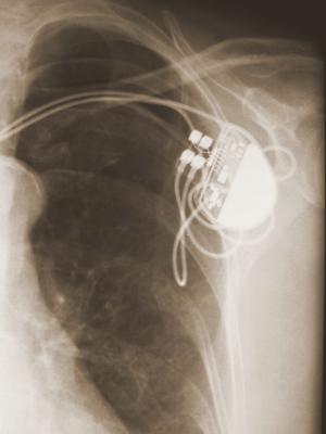
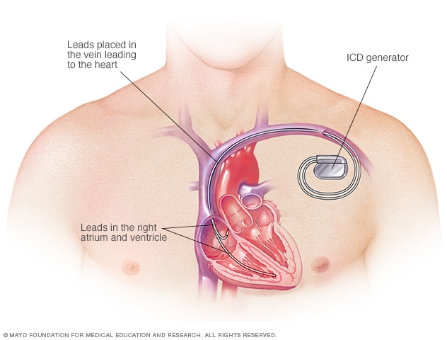
    An ICD is surgically placed under your skin, usually below your left collarbone. One or more flexible, insulated wires (leads) run from the ICD through your veins to your heart.
    Because the ICD constantly monitors for abnormal heart rhythms and instantly tries to correct them, it helps treat cardiac arrest, even when you are far from the nearest hospital.

    How an ICD works

    When you have a rapid heartbeat, the wires from your heart to the device transmit signals to the ICD, which sends electrical pulses to regulate your heartbeat. Depending on the problem with your heartbeat, your ICD could be programmed for the following therapies:
    • Low-energy pacing therapy. You may feel nothing or a painless fluttering in your chest when your ICD responds to mild disruptions in your heartbeat.
    • Cardioversion therapy. A higher energy shock is delivered for a more serious heart rhythm problem. It may feel as if you're being thumped in the chest.
    • Defibrillation therapy. This is the strongest form of electrical therapy used to restore a normal heartbeat. During this therapy, it may feel as if you're being kicked in the chest, and it might knock you off your feet.
      The pain from this therapy usually lasts only a second. There should be no discomfort after the shock ends.
    Usually, only one shock is needed to restore a normal heartbeat. Sometimes, however, you might have two or more shocks during a 24-hour period. Frequent shocks in a short time period are known as ICD storms. If you have ICD storms, you should seek emergency care to see if your ICD is working properly or if you have a problem that's making your heart beat abnormally.
    If necessary, the ICD can be adjusted to reduce the number and frequency of shocks. You may need additional medications to make your heart beat regularly and decrease the chance of an ICD storm.
    An ICD can also be programmed to perform other functions, which include:
    • Antitachycardia (tak-ih-KAHR-dee-uh). If you have an unusually fast heart rate, the ICD delivers painless, low-energy impulses that pace or stimulate the heart to beat normally. This can prevent the need for cardioversion or defibrillation.
    • Pacemaker. Most modern ICDs can also function as a pacemaker, delivering low-energy impulses that stimulate the heart to beat normally.
    • Recording heart activity. The ICD records information about variations in your heart's electrical activity and rhythm. This information helps your doctor evaluate your heart rhythm problem and, if necessary, reprogram your ICD.
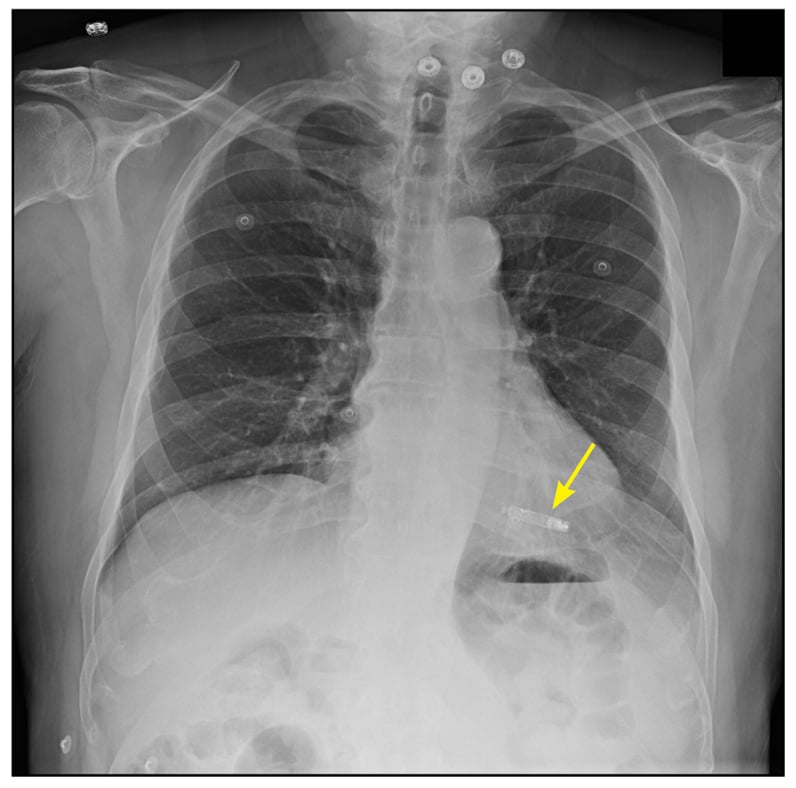
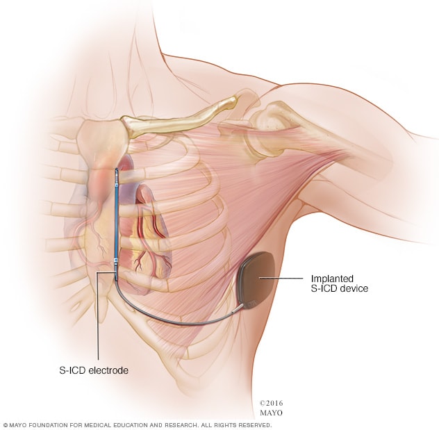
    Subcutaneous ICD

    A subcutaneous ICD (S-ICD), a newer type of ICD, is available at some surgical centers. An S-ICD is implanted under the skin at the side of the chest below the armpit. It's attached to an electrode that runs along your breastbone. You may be a candidate for this device if you have structural defects in your heart that prevent attaching wires to the heart your blood vessels, or if you have other reasons for wanting to avoid traditional ICDs.
    Implanting a subcutaneous ICD is less invasive than an ICD that attaches to the heart, but the device is larger in size than an ICD.

    Who needs an ICD

    You're a candidate for an ICD if you've had sustained ventricular tachycardia, survived a cardiac arrest, or fainted from a ventricular arrhythmia. You might also benefit from an ICD if you have:
    • A history of coronary artery disease and heart attack that has weakened your heart.
    • A heart condition that involves abnormal heart muscle, such as enlarged (dilated cardiomyopathy) or thickened (hypertrophic cardiomyopathy) heart muscle.
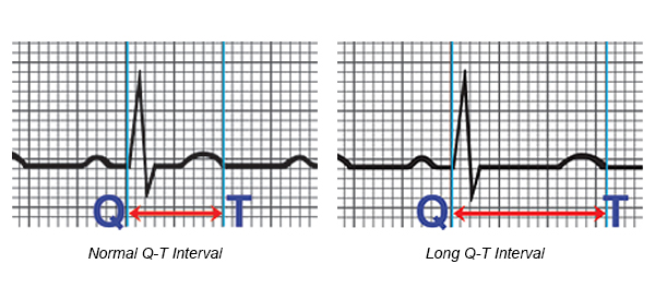
    • An inherited heart defect that makes your heart beat abnormally. These include long QT syndrome, which can cause ventricular fibrillation and death even in young people with no signs or symptoms of heart problems.
    • Having other rare conditions such as Brugada syndrome and arrhythmogenic right ventricular dysplasia also may mean you need an ICD.

    Risks

    Risks associated with ICD implantation are uncommon but may include:
    • Infection at the implant site
    • Allergic reaction to the medications used during the procedure
    • Swelling, bleeding or bruising where your ICD was implanted
    • Damage to the vein where your ICD leads are placed
    • Bleeding around your heart, which can be life-threatening
    • Blood leaking through the heart valve where the ICD lead is placed
    • Collapsed lung (pneumothorax)

    How you prepare

    To determine whether you need an ICD, your doctor might perform a variety of diagnostic tests, which may include:
    • Electrocardiography (ECG). In this noninvasive test, sensor pads with wires attached (electrodes) are placed on your chest and sometimes limbs to measure your heart's electrical impulses. Your heart's beating pattern can offer clues to the type of irregular heartbeat you have.
    • Echocardiography. This noninvasive test uses harmless sound waves that allow your doctor to see your heart without making an incision. During the procedure, a small, plastic instrument called a transducer is placed on your chest.
      It collects reflected sound waves (echoes) from your heart and transmits them to a machine that uses the sound wave patterns to compose images of your beating heart on a monitor. These images show how well your heart is functioning, and recorded images allow your doctor to measure the size and thickness of your heart muscle.
    • Holter monitoring. Also known as an ambulatory electrocardiogram monitor, a Holter monitor records your heart rhythm for 24 hours. Wires from electrodes on your chest go to a battery-operated recording device carried in your pocket or worn on a belt or shoulder strap.
      While wearing the monitor, you'll keep a diary of your activities and symptoms. Your doctor will compare the diary with the electrical recordings and try to figure out the cause of your symptoms.
    • Event recorder. Your doctor might ask you to wear a pager-sized device that records your heart activity for more than 24 hours. Unlike a Holter monitor, it doesn't operate continuously — you turn it on when you feel your heart is beating abnormally.
    • Electrophysiology study (EPS). Electrodes are guided through blood vessels to your heart and used to test the function of your heart's electrical system. This can identify whether you have or might develop heart rhythm problems.
    It's likely you'll be asked not to eat or drink for at least eight hours before your surgery. Talk to your doctor about the medications you take and whether you should continue to take them before your procedure to implant an ICD.

    What you can expect

    During the procedure

    Usually, the procedure to implant an ICD can be performed with numbing medication and a sedative that relaxes you but allows you to remain aware of your surroundings. In some cases, general anesthesia, which puts you to sleep, may be used.
    The procedure usually takes a few hours. During surgery, one or more flexible, insulated wires (leads) are inserted into veins near your collarbone and guided, with the help of X-ray images, to your heart. The ends of the leads are secured to your heart, while the other ends are attached to the generator, which is usually implanted under the skin beneath your collarbone.
    Once the ICD is in place, your doctor will test it and program it for your heart rhythm problem. Testing the ICD might require speeding up your heart and then shocking it back into normal rhythm.

    After the procedure

    You'll stay in the hospital one or two days, and the ICD might be tested once more before you're discharged. Additional testing of your ICD usually doesn't require surgery.

    Treating pain after your procedure

    After surgery, you may have some pain in the incision area, which can remain swollen and tender for a few days or weeks. Your doctor might prescribe pain medication. As your pain lessens, you can take nonaspirin pain relievers, such as acetaminophen (Tylenol, others) or ibuprofen (Advil, Motrin IB, others).
    Unless your doctor instructs you to do so, don't take pain medication containing aspirin because it can increase bleeding risk.
    When you're released from the hospital, you'll need to arrange for a ride home because you won't be able to drive for at least a week.

    Results

    ICDs have become standard treatment for anyone who has survived cardiac arrest, and they're increasingly used in people at high risk of sudden cardiac arrest. An ICD lowers your risk of sudden death from cardiac arrest more than medication alone.
    Although the electrical shocks can be unsettling, there is evidence that the ICD is effectively treating your heart rhythm problem and protecting you from sudden death. Talk to your doctor about how to best care for your ICD.
    After the procedure, you'll need to take some precautions to avoid injuries and make sure your ICD works properly.

    Short-term precautions

    You'll likely be able to return to normal activities such as exercise, work and sex soon after you recover from surgery. Follow your doctor's instructions. For four weeks after surgery, your doctor might ask you to refrain from:
    • Vigorous above-the-shoulder activities or exercises, including golf, tennis, swimming, bicycling, bowling or vacuuming
    • Lifting anything weighing more than 5 pounds
    • Playing contact sports
    • Strenuous exercise programs

    Long-term precautions

    Problems with your ICD due to electrical interference are rare. Still, take precautions with the following:
    • Cellular phones and other mobile devices. It's safe to talk on a cellphone, but avoid placing your cellphone within 6 inches (about 15 centimeters) of your ICD implantation site when the phone is turned on. Although unlikely, your ICD could mistake a cellphone's signal for a heartbeat and slow your heartbeat, causing symptoms such as sudden fatigue.
    • Security systems. After surgery, you'll receive a card that says you have an ICD. Show your card to airport personnel because the ICD may set off airport security alarms.
      Also, hand-held metal detectors often contain a magnet that can interfere with your ICD. Limit scanning with a hand-held detector to less than 30 seconds over the site of your ICD or make a request for a manual search.
    • Medical equipment. Let doctors, medical technicians and dentists you see know you have an ICD. Some procedures, such as magnetic resonance imaging (MRI), magnetic resonance angiography (MRA), and radiofrequency or microwave ablation are not recommended if you have an ICD.
    • Power generators. Stand at least 2 feet (0.6 meters) from welding equipment, high-voltage transformers or motor-generator systems. If you work around such equipment, your doctor can arrange a test in your workplace to see if the equipment affects your ICD.
    • MP3 player headphones. Although the player itself poses little risk, the headphones may be a problem. Most contain a magnetic substance and can interfere with your ICD. Keep your headphones at least 6 inches (about 15 centimeters) from your ICD.
    • Magnets. These might affect your ICD, so it's a good idea to keep magnets at least 6 inches (15 centimeters) from your ICD site.
    Devices that pose little or no risk to your ICD include microwave ovens, televisions and remote controls, AM/FM radios, toasters, electric blankets, electric shavers and electric drills, computers, scanners, printers, and GPS devices.

    Driving restrictions

    If you have an ICD to treat ventricular arrhythmia, driving a vehicle presents a challenge. The combination of arrhythmia and shocks from your ICD can cause fainting, which would be dangerous while you're driving.
    The American Heart Association's guidelines advise avoiding driving for one week after ICD placement, but talk to your doctor for specific recommendations. The guidelines discourage driving during the first six months after your procedure if your ICD was implanted due to a previous cardiac arrest or ventricular arrhythmia.
    If you have no shocks during this period, you'll likely be able to drive again. But if you then have a shock, with or without fainting, tell your doctor and follow his or her recommendations. In most cases, you'll need to stop driving until you've been shock-free for another six months.
    If you have an ICD but have no history of life-threatening arrhythmias, you can usually resume driving within a week after your procedure if you've had no shocks. Discuss your situation with your doctor.
    You usually can't get a commercial driver's license if you have an ICD.

    Battery life

    The lithium battery in your ICD can last up to seven years. The battery will be checked during regular checkups, which should occur every three to six months. When the battery is nearly out of power, your old shock generator is replaced with a new one during a minor outpatient procedure.

    ICDs and end-of-life issues

    If you have an ICD and become terminally ill, your ICD could deliver unnecessary shocks. It's easy to turn off your ICD, and turning it off may prevent unnecessary suffering.
    Talk to your doctor about your wishes. Also talk to family members or another person designated to make medical decisions for you about what you'd like to do in end-of-life care situations.


    Topics

    How to approach Asymptomatic Ventricular Preexcitation (Wolff-Parkinson-White Pattern)

      Tips and Pearls  ·        A significant proportion of patients with ventricular preexcitation remain asymptomatic yet at the risk of life-...