Saturday, January 6, 2018

Interventional Electrophysiology and Cardiac Resynchronization Therapy Delivering Electrical Therapies for Heart Failure

Balloon occlusive coronary sinus venography in the left anterior oblique projection with the branches identified.

Right atrial anatomy and relationship to coronary sinus. OS indicates ostium. Reproduced from Ellenbogen KA et al. Clinical Cardiac Pacing, Defibrillation and Resynchronization Therapy. 3rd ed. Philadelphia, Pa: Elsevier, Inc; 2006:685, with permission from Elsevier, Inc. Copyright 2006.

A, the image  was obtained from Cardio-Optic catheter placed in the coronary sinus. The image reveals the takeoff of a side branch on the right and the main coronary sinus on the left. B, The left anterior oblique fluoroscopic position of the catheter. Venography confirms the location of the branch vessel

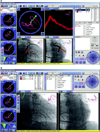
The remote magnetic navigation workstation screen shows the venography and roadmap in the lower 2 panels. B, The same workstation reveals the final lead position.


The top ECG demonstrates right ventricular pacing, and the bottom ECG demonstrates biventricular pacing. Note that with biventricular pacing, an R wave is present in V1 and S wave in lead 1.


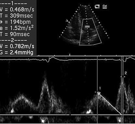
Transmitral Doppler flow reveals the E and A waves.

No comments:

Post a Comment

Topics

How to approach Asymptomatic Ventricular Preexcitation (Wolff-Parkinson-White Pattern)

  Tips and Pearls  ·        A significant proportion of patients with ventricular preexcitation remain asymptomatic yet at the risk of life-...