Simple regional strain pattern analysis to predict response to cardiac resynchronization therapy
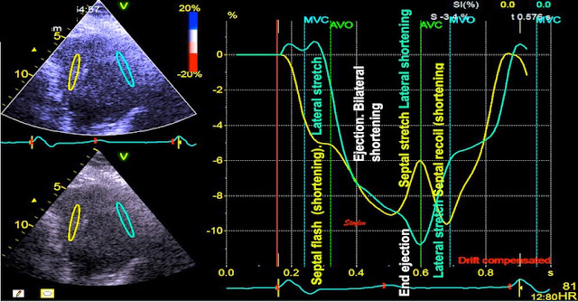
LBBB generates often a classical pattern on echo .The pattern is very distinct in Tissue Doppler of the septum . The classical pattern arises from the time lapse of the activation and relaxation of the two walls, creating a pattern of interaction due to a sequence temporal imbalances of the tension between the two walls.
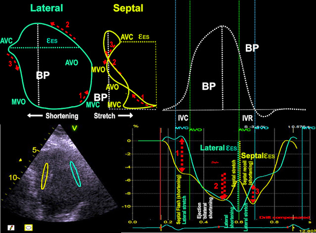
As the septum is activated first, it contracts (shortening - septal flash) without activation of the lateral wall, which stretches. This generates slower pressure build up than a normal IVC, which then is prolonged.
During ejection, the LV volume decreases, so both walls shorten. Tension, however, declines first in the septum, as this was activated first. This leads to a tension imbalance, so the lateral wall continues to shorten, the more relaxed septum stretches.
The stretching of the septum builds up an elastic tension in the septum, which is released as recoil, when tension declines in the lateral wall, showing a post systolic shortening in the septum which is due to elasticity.
This classical pattern is also very evident in the strain curves , and an integrated strain analysis will show this, in a semi quantitative way describing g how work is wasted by looking at the opposing wall .
This classical interaction pattern explains all changes seen in apical velocities (apical rocking), basal velocity curves, strain and strain rate.
Applying estimated LV pressure do not add information, simply because the pressure curve is the same for both walls, so the different strain-pressure loops arises from plotting different strain curves against the same pressure curve, the differences lie in the strain.
References
https://pubmed.ncbi.nlm.nih.gov/22520537/
https://pubmed.ncbi.nlm.nih.gov/30772230/


No comments:
Post a Comment